applications of nanotechnology in medicine and drugs In recent years, applications in medicine and drugs researchers and medication designers have been giving innovative nanotechnology tactics for improving drug delivery to the central nervous system a rising amount of attention (CNS). The treatment of neurological conditions, including Alzheimer's disease, Parkinson's disease, brain tumors, and stroke, might be significantly impacted by nanotechnology in the near future. The use of nanomaterials in the treatment of central nervous system disorders has been shown to be effective in a number of studies, which is relevant to the topic of neurodegeneration. In this context, nanocarriers have been essential in facilitating the targeted administration of chemotherapeutics, which has led to the effective prevention of disease development in malignant brain tumors. Therefore, the use of these compounds in the treatment of CNS disorders is the most effective use of nanomaterials since it boosts the overall impact of medicine and shows the relevance of nano-therapeutics. This research was carried out in order to assess the evidence on the use of nanotechnology in building drug delivery systems with the capacity to pass through the blood-brain barrier and transport therapeutic agents to the central nervous system, this research was carried out. 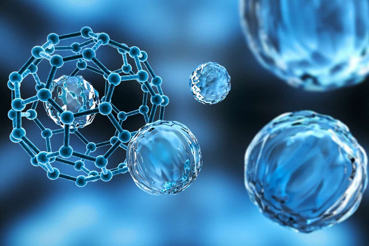 Previously published articles from this issue The following article is in this issue. Keywords Nanotechnology The central nervous system The barrier between the bloodstream and the brain Drug delivery
- The opening statement
Both Alzheimer's and Parkinson's diseases are neurodegenerative conditions that affect a significant number of people. In the United States, stroke is the second most prevalent illness, after Alzheimer's disease, and is linked with neurological disturbance. According to worldwide data, stroke is the third largest cause of death and the second most common disease overall. According to the available data, the incidence of stroke is almost two times more common in Iran than it is in either Europe or North America. According to the World Health Organization, the prevalence of multiple sclerosis illnesses in Iran has been reported as being between 20 and 60 per 100,000 people.  This information was gathered from several sources. The most important step in the treatment of neurological illnesses is the diffusion of medicines through the BBB. Achieving the best possible treatment results in the fight against neurological illnesses requires an extraordinary goal: the secure, suitable, and focused delivery of medicinal molecules to the central nervous system.
- The barrier that separates the bloodstream from the brain.
A membrane known as the blood–brain barrier (BBB) serves to protect the human brain, which is the most delicate and intricate organ in the body. This barrier is well-suited for the purpose of shielding the neurons of the brain from the damaging and poisonous substances that are present in blood. It also has an effect on the diffusion of drugs into the brain tissue. Strong barriers, which prohibit the transmission of any therapeutic or diagnostic substances or other materials to the central nervous system (CNS), divide the brain from other areas of the body and keep it isolated from other regions. In order to have a conceptual understanding of the physiological features of this BBB, it is necessary to first develop methods for the efficient transport of pharmaceuticals and to conduct molecular imaging. The BBB is a barrier that divides the brain from the rest of the circulatory system. The human brain has something in the neighborhood of 100 billion neurons.  There is a possibility that the capillaries in the brain have a diameter of just 7–10 m. The BBB does not have intracellular or valvar gaps, and there is a very narrow opening through which substances may pass from the outside environment to the brain. However, nanoparticles (NPs) paired with polymer coatings may circumvent the BBB and make it easier for medications to be delivered to the central nervous system. The development of this technology has led to a multitude of advancements in the detection and treatment of nervous system illnesses, brain tumors, and head injuries. There are a variety of applicable therapeutic polymers that are now being investigated for clinical usage in the development of novel NPs-based techniques and tactics focused on medication delivery to the brain. These polymers are being used in the treatment of cancer as well as other disorders.
- The movement of substances through the barrier that separates the blood from the brain
Because of the existence of the blood-brain barrier (BBB), which restricts entrance into the brain and protects it from hazardous chemicals as well as infections, pharmaceutical compounds may not be able to penetrate the brain while they are circulating in the blood.  Even very tiny molecules are unable to make their way into the brain because of the barrier. Despite the fact that many necessary molecules are capable of diffusing through this barrier, it often prevents the passage of therapeutic compounds due to the unique characteristics of these compounds. The fact that medications are unable to pass through the BBB underscores the need to adopt NP-based techniques that are specifically aimed at drug delivery. The BBB allows for the diffusion of a restricted fraction of chemicals. These nanocompounds are able to traverse this barrier and make their way to neurons through a variety of pathways. The BBB has been breached using a variety of different NPs. In order for the medicine to cross the blood-brain barrier, the NPs must first break past the tight junctions that are found between the endothelial cells that line the capillaries. Endocytosis and transcytosis of NPs may also make it easier for drugs to move across the layer of endothelial cells. Due to the lipophilic properties of lipid nanoparticles, they are able to penetrate the BBB and make their way into the brain through a variety of different transport channels, such as the paracellular pathway, the transcellular pathway, transcytosis, and receptor-mediated endocytosis.
nanotechnology in medicine and drugs
Nanotechnology in drugs, the controlled manipulation of matter on the atomic and molecular scale to create materials with innovative and remarkably different properties, is a rapidly expanding area of research that holds great potential for many sectors, including medicine, drug construction, and electronics. Medication delivery, gene therapy, diagnostics, and other fields of medical study and practice stand to benefit greatly from this breakthrough. This article does not attempt to give exhaustive coverage of the topic, but rather offers a glimpse into the ways in which nanotechnology may one day impact the practice of medicine, both in the laboratory and the hospital. A Definition of Nanotechnology A Greek word meaning "dwarf" is the origin of the prefix "nano." A nanometer (nm) is one billionth of a meter, or 0.000000001 meters, and is based on the scientific notation of a billionth (10 to the minus 9). The width of a single atom is around three to five nanometers, making a nanometer approximately 40,000 times smaller than a human hair. The typical size of a virus is 100 nm. When applied to medicine, the ability to regulate structures and features at the nanoscale is akin to having a sub-microscopic lab bench on which to work with cell components, viruses, or DNA fragments, utilizing a wide range of tiny tools, robots, and tubes. 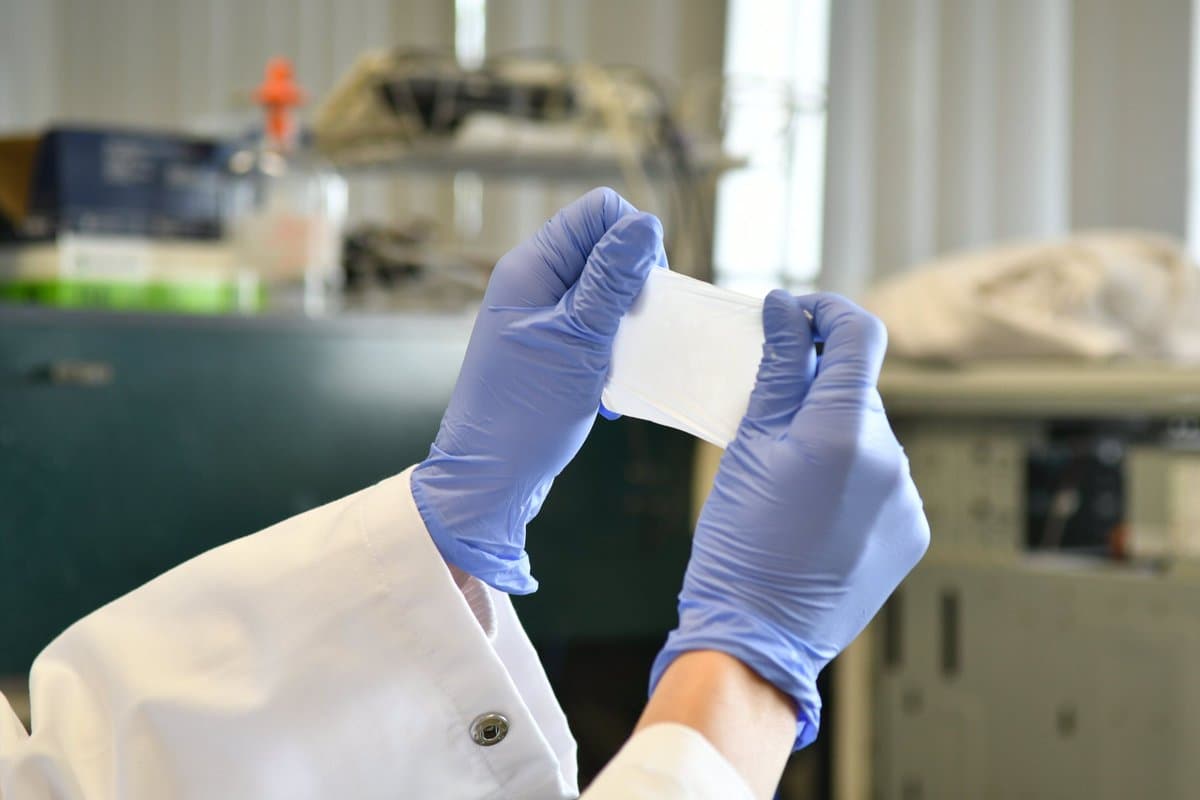 Manipulation of DNA Modifying certain genes or the molecular pathways that control their expression as a therapeutic strategy is gaining popularity. The ability to tailor treatments to the unique genetic makeup of individual individuals is a highly sought-after outcome of this research. As a result, scientists need tools to facilitate their experimentation and the development of potential solutions. The ability to manipulate DNA segments by stretching them out like spaghetti, or developing nanorobots that can "walk" and repair cellular components, are only two examples. That scientific ideal is becoming a reality thanks to nanotechnology. One such study examined the interactions of certain binding proteins by attaching coated latex beads to the ends of modified DNA and then stretching out the DNA strand in an "optical trap" made of a focused beam of light to keep the beads in place. Introducing: Nanobots and Nanostars Meanwhile, nanoscale robots constructed of DNA fragments and operated by a team of researchers at New York University (NYU) can move with the help of two legs under 10 nm in length. Nano Letters presented their study from 2004 detailing how the "nanowalker" uses psoralen molecules attached to the tips of its feet to take its initial steps, two forward and two back.
  One of the researchers, Ned Seeman, thinks it would be possible to construct a molecular-scale production line in which a molecule is pushed until it reaches the appropriate place and a nanobot does some chemical on it. NYU professor Seeman's lab is using DNA nanotechnology to develop a biochip computer and investigate the elusive process of molecular crystallization in biology. The work by Seeman and colleagues exemplifies "biomimetics," the use of nanotechnology to model aspects of biological processes seen in nature, such as DNA behavior, with the goal of developing novel and, perhaps, superior methods. DNA-based nanobots are also being used to kill cancer cells. In order to transport a molecular payload, researchers at Harvard Medical School in the United States recently disclosed in Science how they constructed a DNA "origami nanorobot." The barrel shape of the nanobot suggests that it may carry chemicals that instruct cells to behave in a specific way. Researchers showed they could effectively transport chemicals known to kill leukemia and lymphoma cells. Nanobots are being made with a wide variety of materials. Using gold, for instance, researchers at Northwestern University have developed "nanostars"—simple, specialized, star-shaped nanoparticles that may transport drugs directly to the nucleus of cancer cells. New research published in the journal ACS Nano describes how drug-loaded nanostars act like hitchhikers, being attracted to an over-expressed protein on the surface of human cervical and ovarian cancer cells and depositing their payload directly into the nucleus of those cells.
  The researchers found that by giving their nanobot a star shape, they were able to overcome an issue with using nanoparticles to transport pharmaceuticals: the nanobots were able to release the medications more or less exactly as intended. They claim that the design aids in pinpointing the medication's delivery through laser pulses. On-site drug manufacturing nanofactories Scientists are finding that protein-based medicines may be particularly effective since they can be tailored to deliver specific instructions to cells. However, the problem with conventional methods of drug administration is that the body breaks down most of them before they can do any good. What if, though, such drugs could be produced just where they're needed? Nano Letters published an article by MIT researchers showing how this may be done. In this proof-of-principle work, they show that it is possible for "nanofactories" to self-assemble and create protein molecules on demand at specific places. Nanoparticles designed to express green fluorescent protein (GFP) or luciferase in response to ultraviolet light have been successfully tested in mice. The researchers at MIT conceived the idea while searching for a treatment for metastatic tumors, which are cancer cells that have migrated to other parts of the body.
  More than 90% of cancer deaths are caused by metastatic disease. Scientists are currently investigating this promising new field in order to find new ways to activate nanoparticles that may one day be used as cancer treatments. Fibers with a diameter of less than one thousand nanometers are called nanofibers. Wound dressings, surgical textiles, implants, tissue engineering, and artificial organ components are just some of the many applications for specialty medical materials. The use of carbon nanofibers in scientific instruments and medical imaging has also shown promising results. There are, however, significant challenges to overcome, not the least of which is ensuring that they are consistently the right size. In the past, this process has been time-consuming and costly. Last year, however, scientists at North Carolina State University revealed that they had developed a revolutionary method for making carbon nanofibers with precisely controlled dimensions. In March 2011, they reported in the journal ACS Applied Materials & Interfaces how they used nickel nanoparticles coated with a shell composed of ligands, small organic molecules having functional components that attach directly to metals.
 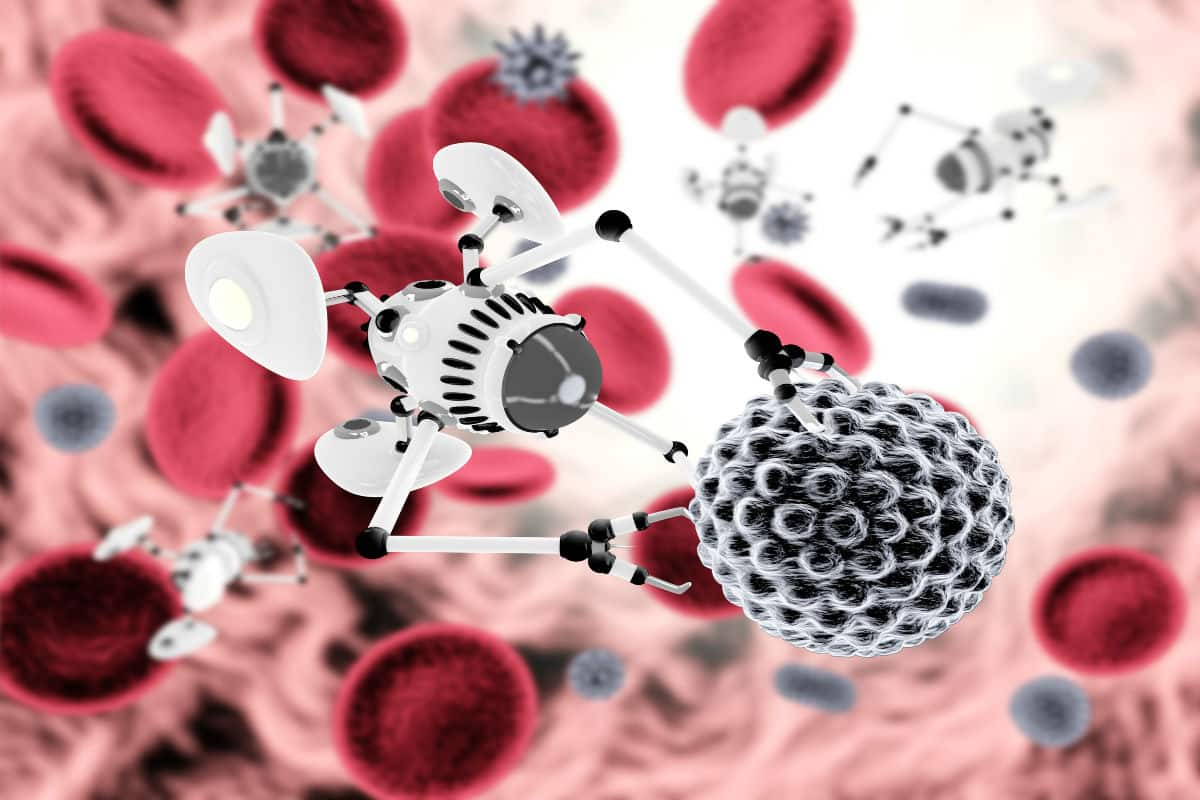 Particularly exciting are nickel nanoparticles because, at elevated temperatures, they facilitate the growth of carbon nanofibers. The researchers also found another benefit to using these nanoparticles: by strategically placing the nanoparticles, they could control where the nanofibers grew, allowing them to produce nanofibers with the ordered precision that is crucial for functional nanoscale materials. Similarly, a neurosurgeon-in-training is employing lead since it is effective as a nanofiber. Matthew MacEwan, a Washington University medical student and entrepreneur, founded a nanomedicine company to improve surgical mesh used around the world. MacEwan speculates that the principal product, a synthetic polymer composed of nanofibers, might also be utilized to treat hernias, fistulas, and other conditions, despite its original intent of treating brain and spinal cord damage. Today, strong, hard surgical meshes are used to repair the membrane that normally shields the brain and spinal cord. MacEwan claims that the lead nanofiber mesh is superior to other mesh materials because it is thinner, more flexible, and more easily incorporated into the patient's own tissues. Each individual thread of nanofiber mesh is countless times smaller than the width of a single cell. The nanofiber material will naturally disintegrate over time, so its use will not only facilitate treatments but also lessen post-op issues for patients.
  Polytechnic Institute of New York University (NYU-Poly) researchers have developed a new approach to synthesizing protein nanofibers. The researchers believe they stumbled upon their finding almost by accident. They were looking at certain cylinder-shaped proteins made from cartilage when they realized that, when present in large enough quantities, some of these proteins spontaneously gathered together and self-assembled into nanofibers. Further research revealed that the synthesis, structure, and attachment of fibers to small molecules could be controlled by adding metal-recognizing amino acids and specific metals. For instance, when nickel was added, the fibers clumped together to form mats that might be used to set off the release of a drug molecule. The researchers believe that their innovative approach will greatly enhance the administration of drugs for treating cancer, cardiovascular disease, and Alzheimer's. They foresee applications in the repair of human organs and the creation of more efficient and compact microprocessors for use in electronics.